
A Beginner's Guide to AI Drug Testing: How 3D Tumors Are Reshaping Medicine
This week, researchers revealed a breakthrough combining 3D-printed patient tumors with real-time AI observation, eliminating the guesswork from cancer treatments.
For decades, one of the most grueling aspects of cancer treatment has been the painful game of trial and error. A patient is diagnosed, a standard chemotherapy or immunotherapy regimen is prescribed, and then the waiting begins. It can take weeks or even months to determine if a specific drug is shrinking the tumor—or if the cancer is simply resisting the treatment while the patient suffers debilitating side effects. But this week, a remarkable medical breakthrough has emerged that promises to eliminate this guesswork entirely.
In mid-August 2026, the intersection of artificial intelligence and biotechnology has reached a profound new milestone. Rather than testing toxic chemicals on a human body to see what works, doctors are leveraging advanced AI vision models to test thousands of therapies on a patient's exact biology in a laboratory setting. This isn't theoretical; it is happening right now, signaling a massive shift toward hyper-personalized oncology.
The Trial-and-Error Problem in Modern Oncology
To understand why this week's developments are so revolutionary, it helps to look at how cancer drugs are traditionally assigned. When a tumor is discovered, doctors rely on clinical guidelines based on population averages. If a patient has a specific type of breast cancer or lung cancer, they are given the first-line treatment that worked for the majority of people in past clinical trials.
However, cancer is notoriously individual. A drug that eradicates a tumor in one patient might be completely ineffective in another, even if their diagnoses look identical on paper. Every single tumor has a unique genetic makeup, a unique microenvironment, and a unique way of fighting off chemical intruders. Until now, finding the precise chemical key to unlock a specific patient's cellular locks was a terrifying race against time.
How Researchers Are Rewriting the Rules This Week
In the past few days, a stunning new methodology has captured the attention of the medical community. The process combines bioprinting with advanced machine learning in a way that sounds like pure science fiction. UCLA researchers have pioneered a technique where they 3D print thousands of mini tumor replicas from a patient's own cancer cells, then let AI watch them all fight drugs in real time, with absolutely no dye needed.
By cultivating these tiny "tumoroids" in a lab, doctors effectively create thousands of biological clones of a patient's disease. They can then bombard these clones with every available cancer drug, in various combinations and dosages, simultaneously. The patient's actual body remains untouched by these experimental combinations, spared from the toxicity of ineffective treatments.
A Beginner's Breakdown of the Process
If you are new to the world of medical AI, the technology stack behind this breakthrough can seem overwhelmingly complex. However, the workflow can be broken down into four clear, sequential steps:
- Step 1: The Biopsy. Doctors extract a small sample of live cancer cells directly from the patient's actual tumor during a standard biopsy or surgical procedure.
- Step 2: 3D Bioprinting. Using specialized 3D bioprinters, researchers multiply these cells into thousands of micro-tumors. These are not flat petri-dish cultures; they are three-dimensional spheres that mimic the exact architecture, density, and behavior of the real tumor inside the patient.
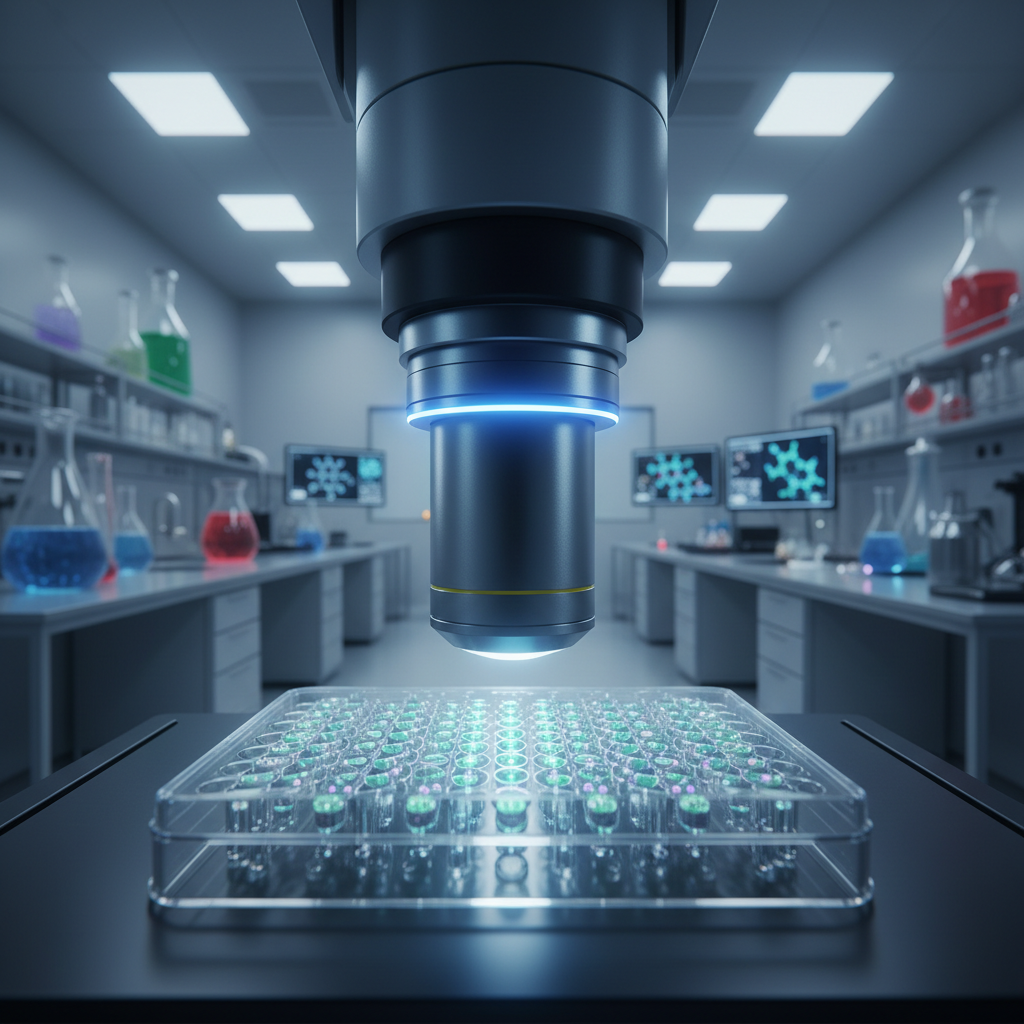
- Step 3: The Chemical Onslaught. Thousands of tiny wells, each containing a micro-tumor, are injected with different cancer drugs. This creates a massive, parallel testing ground.
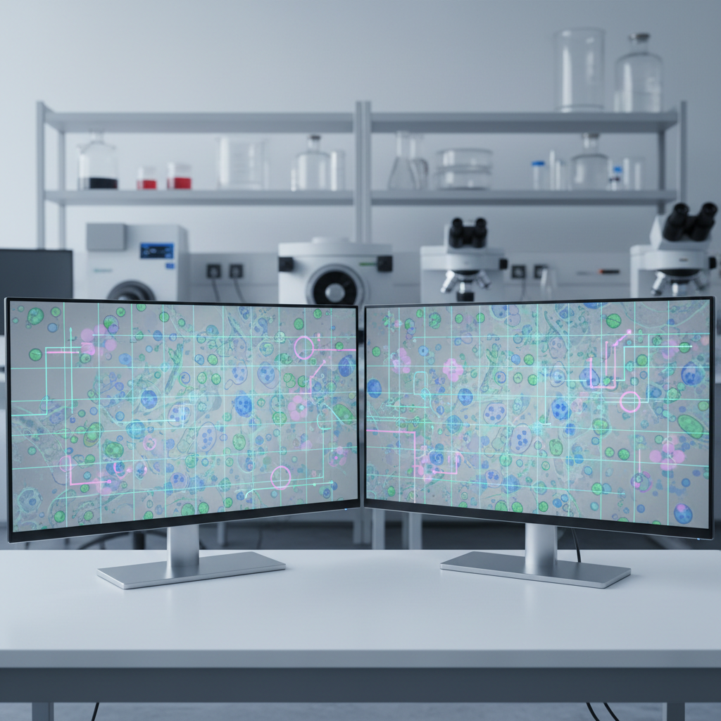
- Step 4: AI Computer Vision Observation. This is where the artificial intelligence takes over. High-resolution cameras record the micro-tumors, and custom AI models watch the footage in real-time, analyzing cellular behavior at a microscopic level to see which tumors die and which survive.
Why "No Dye Needed" is a Massive Leap Forward
You might be wondering: Why do we need AI for this? Couldn't a human scientist just look through a microscope to see if the cells died?
The scale of the data is far too massive for human eyes. Tracking the minute, frame-by-frame structural degradation of thousands of microscopic tumors simultaneously is an impossible task for a team of biologists. In the past, scientists circumvented this by injecting fluorescent chemical dyes into the samples to illuminate cell death. If the well glowed green, the cells were dying.
The problem with chemical dyes is that they are inherently disruptive. Introducing a foreign chemical into a micro-tumor alters its natural biological environment, which can skew the drug test results. The breakthrough this week is that the AI model is so sophisticated—trained on petabytes of microscopic video data—that it can natively identify cell death just by looking at the raw, unaltered footage. It doesn't need the fluorescent crutch. It sees the microscopic warping, the membrane collapses, and the subtle shifts in cellular geometry that indicate the drug is working.
"By removing the chemical dyes, we are getting a pure, unadulterated look at how a patient's specific cancer reacts to pharmacology. The AI acts as an omniscient, hyper-vigilant observer."
The Infrastructure Powering the Cure
The sheer amount of video data generated by watching thousands of micro-tumors in high definition, 24 hours a day, is staggering. This isn't something that can be processed on a standard hospital laptop. It requires immense, localized compute power.
This medical revolution is deeply intertwined with broader technological shifts happening right now. As geopolitical and infrastructural realities push tech giants to reshore their capabilities, investments in supercomputer and chip production are directly enabling hospitals to deploy these heavy vision models on-site. The high-bandwidth, low-latency processing required to interpret live cellular destruction relies entirely on the latest generation of AI infrastructure.
From Mathematics to Human Biology
This week's tumor-tracking development is a perfect example of how foundational AI improvements cascade into real-world applications. When leading AI labs release models capable of deeper logic, the underlying architecture eventually makes its way into specialized fields.
We have recently seen new foundational models utterly redefining AI's reasoning limits by solving previously unbreakable mathematical proofs. That exact same leap in algorithmic reasoning—the ability to look at a complex, chaotic dataset and extract an invisible pattern—is what allows medical AI to look at a chaotic cluster of cancer cells and accurately predict its biological trajectory. Mathematics and biology are converging through the lens of artificial intelligence.
What This Means for Patients Today
For decades, the phrase "personalized medicine" was little more than a marketing buzzword. It usually just meant running a generic DNA panel. Today, as we close out the summer of 2026, personalized medicine actually means cloning your exact disease and forcing an AI to figure out how to destroy it before you take a single pill.
While this 3D-printing and AI-monitoring process is currently rolling out in leading research hospitals and specialized oncology centers, the cost of both bioprinting and AI compute is dropping rapidly. Within the next few years, this could become the standard of care for every newly diagnosed cancer patient. We are moving from a medical system that asks "What usually works?" to a system that definitively answers "What will work for you?"—and that is a shift that will save millions of lives.
Frequently asked questions
What is a 3D printed mini-tumor?
A mini-tumor, or 'tumoroid', is a tiny, three-dimensional cluster of live cancer cells cultivated in a lab from a patient's actual biopsy. It mimics the behavior and structure of the real tumor inside the patient's body.
How does AI help in testing cancer drugs?
AI uses advanced computer vision to watch thousands of these mini-tumors simultaneously in real time. It analyzes microscopic changes to determine exactly which drugs are successfully killing the cancer cells.
Why is it important that the AI doesn't need chemical dyes?
Historically, scientists used fluorescent dyes to see if cells were dying, but these dyes could alter the tumor's natural environment and skew test results. AI vision is advanced enough to detect cell death naturally, ensuring the drug test results are accurate and untainted.
When will this technology be widely available?
As of late 2026, it is currently being utilized in leading research institutions and specialized oncology centers. As the cost of AI compute and 3D bioprinting continues to drop, it is expected to become a standard of care in the coming years.
Join 45,000+ AI builders.
Three tools, two insights, one strategy — every Sunday. The signal cuts through the noise.
Free forever · unsubscribe anytime · no account required
Related reads
Nightshade Explained: How Artists Are Poisoning AI Models in Self-Defense
As AI image generators continue to scrape the web without consent, artists are fighting back with Nightshade—a controversial tool designed to deliberately break AI models from the inside out.

A Beginner's Guide to AlphaFold 3: AI's Medical Revolution
Google DeepMind's AlphaFold 3 is fundamentally changing how scientists create new medications. This beginner-friendly guide explains the massive medical breakthrough.